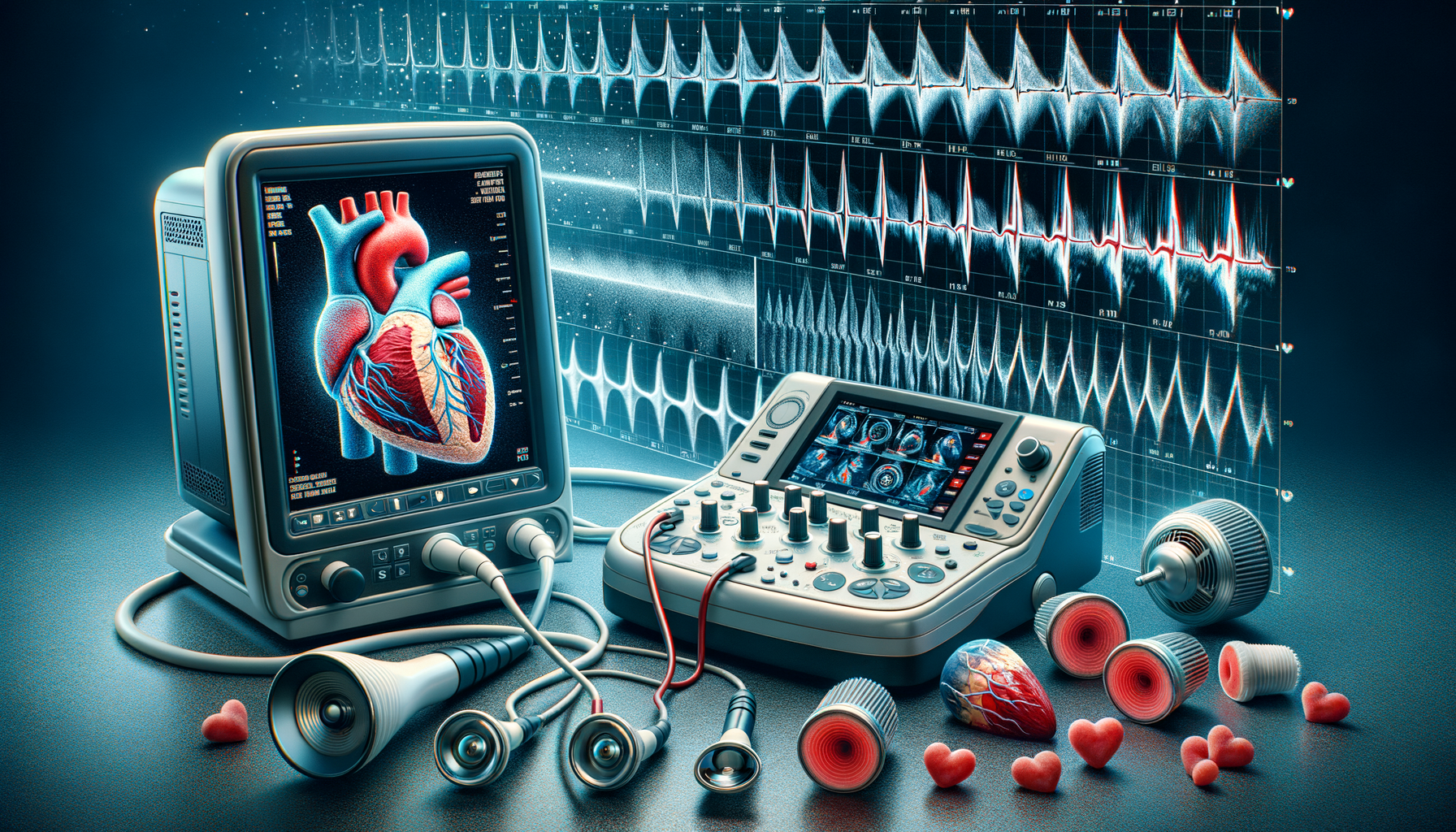
Introduction to Echocardiograms
The heart, a marvel of biological engineering, beats relentlessly to sustain life. Understanding its functionality is crucial, especially when diagnosing cardiovascular conditions. Enter the echocardiogram, a non-invasive diagnostic tool that uses ultrasound waves to create images of the heart. This remarkable technology allows healthcare professionals to visualize the heart’s structure and function in real-time, offering invaluable insights into its health. Whether it’s assessing heart valve function, detecting heart diseases, or monitoring the effects of heart treatments, echocardiograms play a pivotal role in modern medicine.
The Science Behind Echocardiograms
At the core of an echocardiogram is the use of high-frequency sound waves, known as ultrasound, which are inaudible to the human ear. When these sound waves are directed towards the heart, they bounce back upon hitting different structures, such as the heart walls and valves. A transducer, a device placed on the chest, captures these returning echoes and converts them into electrical signals. These signals are then processed by a computer to create detailed images of the heart. This process not only reveals the heart’s anatomy but also provides dynamic information about how well the heart is pumping and the condition of its valves.
Types of Echocardiograms
Echocardiograms come in several forms, each serving a specific purpose. The most common type is the transthoracic echocardiogram (TTE), which involves placing the transducer on the chest. Another type, the transesophageal echocardiogram (TEE), involves inserting a specialized transducer into the esophagus, offering clearer images of the heart’s structures. Additionally, stress echocardiograms are performed to evaluate the heart’s function under stress, often induced by exercise or medication. Each type of echocardiogram provides unique insights, enabling precise diagnosis and tailored treatment plans.
Clinical Applications and Benefits
Echocardiograms are indispensable in diagnosing a wide range of heart conditions. They help in assessing heart valve function, detecting congenital heart defects, and evaluating the effectiveness of treatments. By providing real-time images, echocardiograms allow clinicians to monitor changes in heart function over time. The procedure is safe, painless, and free of radiation, making it a preferred choice for patients of all ages. Additionally, the ability to perform echocardiograms at the bedside enhances their utility in critical care settings, ensuring timely diagnosis and intervention.
Future Prospects and Innovations
The field of echocardiography is continuously evolving, with technological advancements enhancing its diagnostic capabilities. Innovations such as three-dimensional echocardiography and contrast-enhanced echocardiography are providing even more detailed images of the heart. Additionally, the integration of artificial intelligence is set to revolutionize the interpretation of echocardiograms, offering automated analysis and improved accuracy. As these technologies advance, echocardiograms will continue to be a cornerstone of cardiovascular diagnostics, offering hope and improved outcomes for patients worldwide.
Conclusion: A Heartfelt Insight
In conclusion, echocardiograms stand as a testament to the wonders of medical technology, offering a non-invasive, detailed view of the heart’s inner workings. For patients and healthcare providers alike, this diagnostic tool provides peace of mind and clarity, ensuring that heart conditions are detected and managed with precision. As technology continues to advance, the role of echocardiograms in heart health will only become more significant, guiding us towards a future where heart diseases are diagnosed earlier and treated more effectively.


Leave a Reply