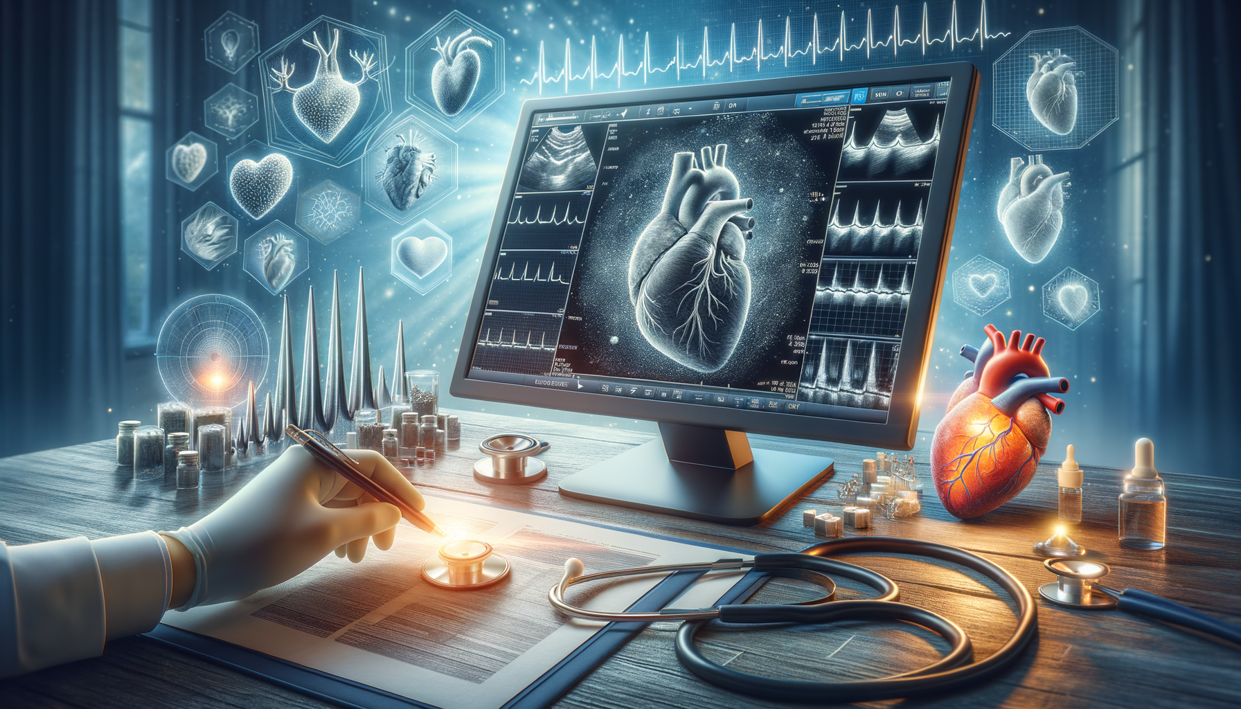
The Basics of Echocardiograms
An echocardiogram, often referred to as an “echo,” is a non-invasive diagnostic tool that uses ultrasound waves to create images of the heart. This technique allows doctors to observe the heart’s structure and function in real time. Unlike other imaging methods, echocardiograms do not involve radiation, making them a safer option for repeated use. The procedure is typically performed by a sonographer and interpreted by a cardiologist.
The primary purpose of an echocardiogram is to evaluate the heart’s chambers and valves. By examining these components, healthcare providers can detect abnormalities such as valve stenosis, regurgitation, or congenital heart defects. Additionally, echocardiograms can assess the heart’s pumping strength, which is vital for diagnosing conditions like heart failure. The test is versatile and can be adapted to various forms, including transthoracic, transesophageal, and stress echocardiograms, each serving specific diagnostic needs.
In summary, echocardiograms are an essential part of cardiac care, providing detailed insights into the heart’s anatomy and function without the risks associated with radiation. Their non-invasive nature and adaptability make them a cornerstone in diagnosing and managing heart conditions.
How Echocardiograms Aid in Diagnosing Heart Conditions
Echocardiograms are pivotal in diagnosing a wide range of heart conditions. One of the key advantages of this imaging technique is its ability to detect heart valve abnormalities. Valves that do not open or close properly can lead to conditions such as aortic stenosis or mitral valve prolapse. By visualizing the heart valves in action, echocardiograms can identify these issues early, allowing for timely intervention.
Another significant role of echocardiograms is in assessing heart muscle function. The echocardiogram can measure the ejection fraction, which indicates how well the heart is pumping blood. A reduced ejection fraction is a hallmark of heart failure, and regular echocardiograms can monitor changes in heart function over time. This is crucial for adjusting treatment plans and improving patient outcomes.
Echocardiograms are also instrumental in detecting congenital heart defects, which are structural problems present at birth. These defects can range from minor issues that require monitoring to significant abnormalities necessitating surgical correction. The ability to visualize these defects in detail makes echocardiograms an invaluable tool in pediatric cardiology.
In essence, echocardiograms provide a comprehensive view of the heart’s structure and function, enabling accurate diagnosis and management of various cardiac conditions. Their role in early detection and ongoing monitoring is indispensable in modern cardiology.
The Role of Echocardiograms in Monitoring Heart Health
Beyond diagnosis, echocardiograms play a crucial role in the ongoing monitoring of heart health. For patients with known cardiac conditions, regular echocardiograms can track the progression of disease and the effectiveness of treatments. This is particularly important in managing chronic conditions like heart failure, where changes in heart function can occur gradually.
For patients who have undergone cardiac surgery, echocardiograms can assess the success of the procedure and monitor for potential complications. For instance, after valve replacement surgery, echocardiograms can ensure that the new valve is functioning correctly and that there are no signs of leakage or obstruction.
In addition to monitoring specific conditions, echocardiograms can be used as a preventive measure in individuals at risk of developing heart disease. By identifying early signs of cardiac dysfunction, such as reduced ejection fraction or abnormal heart wall motion, interventions can be implemented to prevent the progression to more serious conditions.
Overall, the ability to monitor heart health over time makes echocardiograms a vital component of comprehensive cardiac care. They offer a non-invasive, reliable method for assessing heart function and guiding treatment decisions.
Comparing Echocardiograms with Other Cardiac Imaging Techniques
While echocardiograms are a cornerstone in cardiac imaging, they are not the only available technique. Other imaging methods, such as MRI, CT scans, and nuclear imaging, also play roles in cardiac diagnostics. Each method has its strengths and limitations, making them suitable for different clinical scenarios.
Magnetic Resonance Imaging (MRI) provides detailed images of the heart’s structure and is particularly useful in evaluating complex congenital heart defects and cardiac tumors. However, MRI is more expensive and less accessible than echocardiograms, and it is not suitable for patients with certain implants or those who are claustrophobic.
Computed Tomography (CT) scans offer rapid imaging and are excellent for assessing coronary artery disease. They provide a detailed view of the coronary arteries, helping to identify blockages that might lead to heart attacks. However, CT scans involve exposure to radiation, which is a consideration for repeated use.
Nuclear imaging techniques, such as positron emission tomography (PET), provide functional information about the heart, such as blood flow and metabolism. These methods are highly sensitive but also involve radiation exposure and are typically more costly.
In comparison, echocardiograms offer a unique combination of safety, accessibility, and versatility. While other imaging techniques may provide more detailed structural or functional information, echocardiograms remain a first-line tool due to their non-invasive nature and ability to provide real-time insights into heart function.
The Future of Echocardiography in Heart Health
The field of echocardiography is continually evolving, with advancements in technology enhancing its diagnostic capabilities. One promising development is the integration of 3D and 4D imaging, which provides more detailed views of the heart’s structure and function. These advancements allow for better visualization of complex heart conditions and more accurate assessments of heart function.
Artificial intelligence (AI) is another area poised to revolutionize echocardiography. AI algorithms can assist in interpreting echocardiograms, reducing the time required for analysis and increasing diagnostic accuracy. This is particularly beneficial in settings with limited access to experienced cardiologists, as AI can help bridge the gap in expertise.
Portable echocardiography devices are also becoming more prevalent, making it possible to perform heart assessments outside of traditional healthcare settings. This is particularly valuable in remote or underserved areas, where access to advanced medical equipment is limited. Portable devices can facilitate early diagnosis and timely intervention, improving outcomes for patients with heart disease.
As technology continues to advance, echocardiography will remain a vital tool in heart health. Its ability to adapt and integrate new technologies ensures that it will continue to provide valuable insights into cardiac function and contribute to the overall improvement of heart health care.


Leave a Reply